 |
| Sequence I: Basic principles and classification |
This post is helpful for both Beginners and Experts. I hope you Like it. Let's Start.
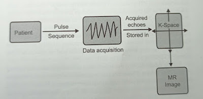
A pulse sequence is an interplay of various parameters leading to a complex cascade of events with RF pulses and gradients to form an MR image (Fig. 1.1).
So basically pulse sequence is a time chart of the interplay of:
- Patient's net longitudinal magnetization
- Transmission of RF pulses (90,180 degrees or any degree)
- X, Y, and Z gradient activation for localization and acquisition of signal (echo)
- K-Space filling with acquired signal or echoes.
 |
| Fig. 1.1: Steps in image acquisition |
Classification of Sequence I in MRI
Pulse sequences can be broadly divided into two categories- spin-echo and gradient-echo sequences. Inversion recovery and echo-planar imaging (EPI) can be applied theoretically to both spin-echo and gradient-echo sequences. However, in practice inversion recovery is applied to spin-echo sequences and EPI is used with gradient-echo sequences. For practical purposes, let's consider the following four types of sequences:
1. Spin-echo sequence (SE)
2. Gradient Echo sequence (GRE)
3. Inversion Recovery sequences (IR)
4. Echo Planar Imaging (EPI).
Spin Echo (SE) Pulse Sequence
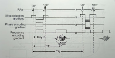
It consists of 90 and 180 degree RF pulses. The excitatory 90-degree pulse flips the net magnetization vector along Z-axis into the transverse (X-Y) plane. The transverse magnetization (TM) precessing at Larmor frequency induces a small signal called free induction decay (FID) in the receiver coil. FID is weak and insufficient for image formation.
Also read: MRI Radiofrequency Coils
Also, the amount of TM magnetization reduces as protons start dephasing. Hence a rephasing 180-degree pulse is sent to bring protons back into the phase. This rephasing increases magnitude of TM and a stronger signal (spin echo) is induced in the receiver coil. This gives the sequence its name. The time between two 90 degree pulses is called TR (Time to Repeat). The time between 90-degree pulse and reception of echo (signal) is TE (Time to Echo) (Fig. 1.2).
For the localization of the signal, the slice selection gradient is turned on when RF is sent. Phase encoding gradient is turned on between excitation (90 degrees) pulse and signal measurement. Phase encoding gradient has different strengths for each TR. Frequency encoding (readout gradient) is turned on during signal measurement.
 |
| Fig. 1.2: Spin-echo (SE) sequence |
SE sequence forms the basis for understanding all other sequences. It is used in almost all examinations. T1-weighted images are useful for demonstrating anatomy. Since the diseased tissues are generally more edematous and or vascular, they appear bright on T2- weighted images. Therefore, T2- weighted images demonstrate pathology well.
Modification of SE Sequences in MRI
In the conventional SE sequence, one line of K-Space is filled per TR. SE sequence can be modified to have more than one echo (line of K-Space) per TR. This is done by sending more than 180 pulses after the excitatory 90-degree pulse. Each 180-degree pulse obtains one echo. Three routinely used modifications in the SE sequence include dual SE (two 180 degree pulses per TR), fast SE (multiple 180-degree pulses per TR), and single-shot fast SE (fast SE with only half of the K-Space filled).
1. Dual spin-echo Sequence
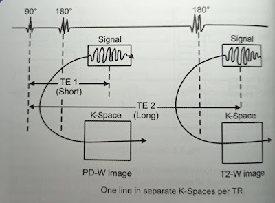
Two 180 degree pulses are sent after each 90-degree pulse to obtain two echoes per TR. The PD + T2 double echo sequence (Fig. 1.3) is an example of this modified sequence. This sequence is run with a long TR. After the first 180 degrees pulse, since TE is short, the image will be proton density-weighted (long TR, short TE). After the second 180 degree pulse, TE will be long giving a T2-W image (long TR, long TE). Both these echoes contribute separate K-Space lines in two different K-Spaces.
 |
| Fig. 1.3: Double Echo sequence |
2. Fast (turbo) spin-echo Sequence
In a fast SE sequence, multiple 180-degree rephasing pulses are sent after each 90-degree pulse. It is also called a multi spin-echo or Turbo spin-echo sequence (Fig. 1.4). In this sequence, multiple echoes are obtained per TR, one echo with each 180-degree pulse. All echoes are used to fill a single K-Space. Since K-Space is filled much faster with multiple echoes in a single TR the scanning speed increases considerably.
Turbo factor: turbo factor is the number of 180-degree pulses sent after each 90-degree pulse. It is also called echo train length. The amplitude of the signal (echo) generated from the multiple refocusing 180-degree pulses varies since the TE goes on increasing. The TE at which the center of the K-Space is filled is called as 'TE effective. The amplitude of the signal is maximum at the TE effective. Short turbo factor decreases effective TE and increases T1 weighting. However, it increases scan time. The long turbo factor increases effective TE, increases T2 weighting, and reduces scan time. Image blurring increases with turbo factor because more echoes obtained at different TE form the same image.
 |
| Fig. 1.4: Fast/Turbo spin-echo sequence |
When a negative 90-degree pulse is sent at the end of the echo train, the magnetization for tissues with high T2 flips quickly back in the longitudinal plane at the end of each TR. This makes fast SE sequence even faster. The fast SE sequences using this 90-degree flip-back pulse at the end of each TR are called fast recovery (FRFSE, GE), driven equilibrium (DRIVE, Philips) or RESTORE (Siemens) Sequence.
3. Single-shot Fast spin-echo Sequence
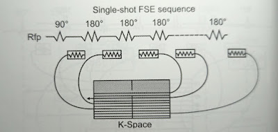
This is a fast SE sequence in which all the echoes required to form an image are acquired in a single TR. Hence it is called 'single-shot sequence. In this sequence, not only all K-Space lines are acquired in a single excitation but also just over a half of the K-Space is filled reducing the scan time further by half (Fig. 1.5). The other half of the K-Space is mathematically calculated with half-Fourier transformation. Thus, for example, to acquire an image with a matrix of 128 x 128 it is sufficient to acquire only 72 K-Space lines. This number can be further reduced by the use of parallel imaging, greatly reducing blurring.
 |
| Fig. 1.5: Single-shot fast-spin echo sequence |
Gradient Echo (GRE) Sequence in MRI
There are basically three differences between SE and GRE sequences.
1. There is no 180-degree pulse in GRE. Rephasing of TM in GRE is done by gradients; Particularly by reversal of the frequency encoding gradient. Since rephasing by gradient gives a signal this sequence is called Gradient echo sequence.
2. The flip angle in GRE is smaller, usually less than 90 degrees. Since the flip angle is smaller there will be the early recovery of longitudinal magnetization (LM) such that TR can be reduced, hence the scanning time.
3. Transverse relaxation can be caused by a combination of two mechanisms
- Irreversible dephasing of TM resulting from nuclear, molecular, and macromolecular magnetic interactions with a proton.
- Dephasing is caused by magnetic field inhomogeneity. In the SE sequence, the dephasing caused by a magnetic field inhomogeneity is eliminated by a 180-degree pulse. Hence there is true transverse relaxation in the SE sequence. In the GRE sequence, dephasing effects of magnetic field inhomogeneity are not compensated, as there is no 180-degree pulse. T2 relaxation in GRE is called T2* (T2 star) relaxation.
1/T2* =1/T2 + 1/relaxation by inhomogeneity Usually T2*<T2.
So the basic GRE sequence is as shown in the diagram (Fig. 1.6).
Types of GRE Sequences in MRI
GRE sequences can be divided into two types depending on what is done with the residual transverse magnetization (TM) after the reception of the signal in each TR. If the residual TM is destroyed by RF pulse or gradient such that it will not interfere with the next TR, the sequences are called spoiled or incoherent GRE sequences. In the second type of the GRE sequences, the residual TM is not destroyed. In fact, it is refocused such that after a few TRs steady magnitude of LM and TM is reached. These sequences are called steady-state or coherent GRE sequences.
 |
| Fig. 1.6: Gradient Echo sequences (GRE). Reversal of frequency encoding gradient (instead of 18-0 degree pulse) causes rephasing of protons after RFp is switched off. TS= time to signal |
SPOILED/INCOHERENT GRE Sequences
These sequences usually provide T1-weighted GRE images (FLASH/ SPGR/T1-FFE). These sequences can be acquired with echo times when water and fat protons are in-phase and out-of-phase with each other. This 'in-and out-of-phase imaging' is used to detect fat in the lesion or organs and is discussed in chapters 6 and 7. Spoiled GRE sequences are modified to have time-of-flight MR Angiographic sequences. The 3D versions of these sequences can be used for dynamic multiphase post-contrast T1-weighted imaging, Examples include 3D FLASH and VIBE (Siemens), LAVA and FAME (GE), and THRIVE (Philips).
STEADY STATE (SS) Sequences
When the residual transverse magnetization is refocused keeping TR shorter than T2 of the tissues, a steady magnitude of LM and TM is established after a few tries. Once the steady-state is reached, two signals are produced in each TR: FID (S+) and spine-echo (S-). Depending on what signal is used to form the images, SS sequences are divided into 3 types.
1. Post-excitation refocused steady-state sequences.
Only FID (S+) component is sampled. Since S+ echo is formed after RF excitation (pulse), it is called post-excitation refocused. e.g. FISP/GRASS/FFE/FAST.
2. Pre-excitation refocused steady-state sequences.
Only spin-echo (S-) component is used for image formation. S-echo is formed just before the next excitation hence the name pre-excitation refocused.
e.g. PSIF (reversed FISP)/SSFP/T2-FFE/CE-FAST.
3. Fully refocused steady-state sequences.
Both FID (S+) and Spin echo (S-) components are used for image formation. These are also called 'balanced-SSFP' sequences as gradients in all three axes are balanced making them motion insensitive.
e.g. True FISP/FIESTA/Balanced FFE
SS sequences have very short TR and TE making them fast sequences that can be acquired with breath-hold. They can be used to study rapid physiologic processes (e.g. events during a cardiac cycle) because of their speed. Balanced-SSFP sequences have the highest possible SNR among all MR sequences and show tissues or structures with high T2 (e.g. fluid) more bright than usual T2-weighted images. However, they lack internal spatial resolution and do not demonstrate gray-white differentiation in the brain or corticomedullary differentiation in the kidneys.
Inversion Recovery (IR) Sequence
IR sequence consists of an inverting 180-degree pulse before the usual spin-echo or gradient-echo sequence. In practice, it is commonly used with SE sequences (Fig. 1.7).
The inverting 180-degree pulse flips LM from the positive side of the Z-axis to the negative side of the Z-axis. This saturates all the tissues. LM then gradually recovers and builds back along the positive side of the Z-axis. This LM recovery is different for different tissues depending on their T1 values. Protons in fat recover faster than the protons in water. After a certain time, the usual sequence of 90-180 degree pulses is applied. Tissues will have different degrees of LM recovery depending on their T1 values. This is reflected in increased T1 contrast in the images.
 |
| Fig. 1.7: Inversion recovery (IR) sequence |
The time between inversion 180 degrees and excitatory 90-degree pulses is called 'time to invert or TI'. TI is the main determinant of contrast in IR sequences.
Why is the inverting 180-degree pulse used? What is achieved? The inversion 180-degree pulse flips LM along the negative side of the Z-axis. This saturates fat and water completely at the beginning. When the 90-degree excitatory pulse is applied after LM has relaxed through the transverse plane, the contrast in the image depends on the amount of longitudinal recovery of the tissues with different T1.
An IR image is more heavily T1-weighted with a large contrast difference between fat and water. Apart from getting heavily T1-weighted images to demonstrate anatomy, IR sequence is also used to suppress particular tissue using different Tl.
Tissue Suppression
At the halfway stage during recovery after a 180-degree inversion pulse, the magnetization will be at zero level with no LM available to flip into the transverse plane. At this stage, if the excitatory 90-degree pulse is applied, TM will not be formed and no signal will be received. If the TI corresponds with the time a particular tissue takes to recover till halfway stage there will not be any signal from that tissue. IR sequences are thus used to suppress certain tissues by using different TI. The Tl required to null the signal from tissue is 0.69 times the T1 relaxation time of that tissue.
Types of IR sequences in MRI
IR sequences are divided based on the value of TI used. IR sequences can be of short, medium or long TI.
Short TI IR sequences use TI in the range of 80-150 ms and an example is STIR. In Medium TI IR sequences, TI ranges from 300 to 1200 ms, and an example is MPRAGE (Siemens). Long Tl ranges from 1500 to 2500 ms and an example is a FLAIR.
SHORT TI (tau) IR Sequence (STIR)
When a 90-degree pulse is applied at short TI, LM for all or virtually all tissues is still on the negative side. The tissues with short T1 have near-zero magnetization, so don't have much signal. Most pathologic tissues have increased T1 as well as T2. Moderately high TE used in STIR allows tissues with high T2 to retain signals while tissues with short T2 will have reduced signal. This results in increased contrast between tissues with short T1-T2 and tissues with long T1-T2. In short TI IR sequences, fat is suppressed since fat has short T1. Most pathologies appear bright on STIR making them easier to pick up.
LONG TI IR Sequence
When the 90-degree pulse is applied at long TI, the LM of most tissues is almost fully recovered. Since water has long T1, its LM recovery is at halfway stage at long Tl. This results in no signal from a fluid such as CSF. The sequence is called fluid-attenuated inversion recovery sequences (FLAIR). In FLAIR, long TE can be used to get heavily T2-weighted without problems from CSF partial volume effects and artifacts as it is nulled. As in STIR, most of the pathologies appear bright on FLAIR.
Echo Planar Imaging (EPI)
Scanning time can be reduced by filling multiple lines of K-Space in a single TR. EPI takes this concept to the extreme. All the lines of K-Space, required to form an image, are filled in a single TR. Since the entire 2D raw data set (i.e. a plane of data or echo) could be filled in single echo decay, the term 'Echo Planar Imaging' was given to this technique by Sir Peter Mansfield in 1977.
Multiple echoes generated in a single TR in EPI are phase-encoded by different slopes of the gradient. These multiple echoes are either generated by 180-degree rephasing pulses or by the gradient. Therefore, EPI can be Spin-Echo EPI (SE-EPI) or Gradient echo EPI (GRE-EPI). However, in SE-EPI multiple 180 degrees, RF pulses cause excessive energy deposition in patient tissues and a long train of 180-degree pulses would take so long time that most of the signal would be lost before satisfactory data could be acquired. Hence SE-EPI is not routinely used. In GRE-EPI, rephasing is done by rapidly switching read-out and phase encoding gradients on and off. This requires gradient strength higher than 20 mT/m.
GRE-EPI is very sensitive to susceptibility artifacts because T2" decay is not compensated in GRE sequences. The signal-to-noise ratio (SNR) is poor in single-shot EPI.
These problems can be overcome to a certain extent by the hybrid sequence. Hybrid sequence combines advantages of gradient (speed) and RF pulses (compensation of T2* effects). SNR can be improved by the use of multi-shot EPI. In multi-shot EPI, only a portion of K-Space is filled in each TR. Multi-shot EPI improves SNR and spatial resolution and gives better PD and T1-weighting at the cost of a slight increase in time.
EPI has revolutionized MR imaging with its speed. EPI has the potential for interventional and real-time MRI. Presently it is used in diffusion imaging, perfusion imaging, functional imaging with BOLD, cardiac imaging, and abdominal imaging.
I hope you understand what are the Basic principles and Classification of Sequence I in MRI.
MRI all sequence explained- This video representation is all about MRI pulse sequence.👇👇🎯

Post a Comment