Ultrasound is the name given to high-frequency sound waves, over 20,000 cycles per second (20kHz). These waves, inaudible to humans, can be transmitted in beams an d are used to scan the tissue of the body.
The ultrasound waves are generated by a piezoelectric transducer which is capable of changing electrical signals into mechanical (ultrasound) waves. The same transducer can also receive the reflected ultrasound and change it back into electrical signals. Transducers are both transmitters and receivers of ultrasound.
Orientation of image (Guideline)
 |
| A finger on the transducer should produce an image on the same side of the screen. If the image is on the wrong side, rotate the transducer by 180° |
It is possible for the images on the monitor to be reversed so that, on traverse scans, the left side of the patient is seen on the right side of the screen. Although there may be an indicator on the transducer, it is essential before scanning (ultrasound) to check visually which side of the transducer produces which side of the image. This is the best done by putting a finger at one end of the transducer and seeing where it appears on the screen. If incorrect, rotate the transducer 180,d and check again. Only longitudinal scans, the head of the patient should be on the left side and the feet on the right side have the screen.
 |
| Two axial images of the same fetal head, but aligned 180° differently. When starting to scan, the images on the screen must be tested as shown in |
Background of the image:
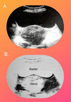
The image on the screen may be predominantly black and predominantly white. It may have a white background with black echoes, or a black background with white echoes showing as spots or lines. There is usually switch to make this change; if not, an engineer should adjust the machine show that is always show a black background with white echoes.
 |
| (a) white echoes on black background (correct) (b) Black echoes on white background (incorrect) [ Transverse images of an enlarged uterus with the background change ] |
Distribution of the ultrasound beam:
Body tissue reflects ultrasound in two different ways. Some tissues act like mirrors, sending the waves directly back. Other scatters the waves in the way fog scatters a beam of light.
e.g. The diaphragm is a "mirror", Known technically as a "specular reflector".
The monitor will show a clear and exact image that corresponds well with the position and the shape of the diaphragm. The liver, however, setters ultrasound waves and the position of the dots shown on the screen does not exactly correspond with any particular detail in the liver. This is an "interference pattern" resulting from waves being scattered in different directions. In either case, the use of a black background With value white echoes permits better differentation.


Post a Comment