 |
| CT- Beginners Tips |
CT protocol for Routine Thorax
Indication
Screening Infection/Inflammation, Trauma, Masses of Lung pleura and mediastinum staging lymphoma lesions of the chest wall and esophagus Followups
Patient positioning
Head First Supine with Arms elevated above the Head
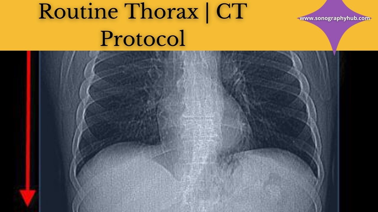
Topogram Position/Landmark
Anteroposterior; 1 inch below the level of the chin to Umbilicus.
Mode of Scanning
Helical with single breath-hold.
Scan Orientation
Caudocranial
- Starting Locations- The imaginary line joining the two costophrenic angles
- End Location- 1 cm above the Apex of the Lung
Gantry Tilt
Nill
Field of view
Just fitting to the Thoracic Cavity including the soft tissues of the Chest wall.
Contrast Administration
Intravenous oral Air/Positive Contrast for Esophageal Evaluation
The volume of Contrast
60-100 mL.
Rate of Injection of Contrast
2-2.5 ml/sec
Scan Delay
35-45 sec
Slice Thickness in Reconstruction
3-5 mm
Slice Interval
1.5-2.5 mm
Reconstruction Algorithm/Kernel
Medium smooth sharp for pulmonary parenchyma and Bone
3D-Reconstructions
- MPR,
- MIP,
- VRT if needed
Comments
Noncontrast Scans should be taken in the region of interest or lesion detected on Chest radiography or Topogram.
When scanning the chest and Abdomen in a single examination Abdomen protocol should be followed
Scanned Volume should be extended to the level of Adrenals in suspected Bronchogenic Cancers and to the level of the Celiac axis in the Carcinoma Esophagus.
Additional Prone and Decubitus Scans should be taken through the region of interest in cases of Cavitary Lesions to demonstrate the mobility of its contents.
Criteria of Good Image Quality
Absence of motion artefacts and Respiratory misregistration.

Post a Comment