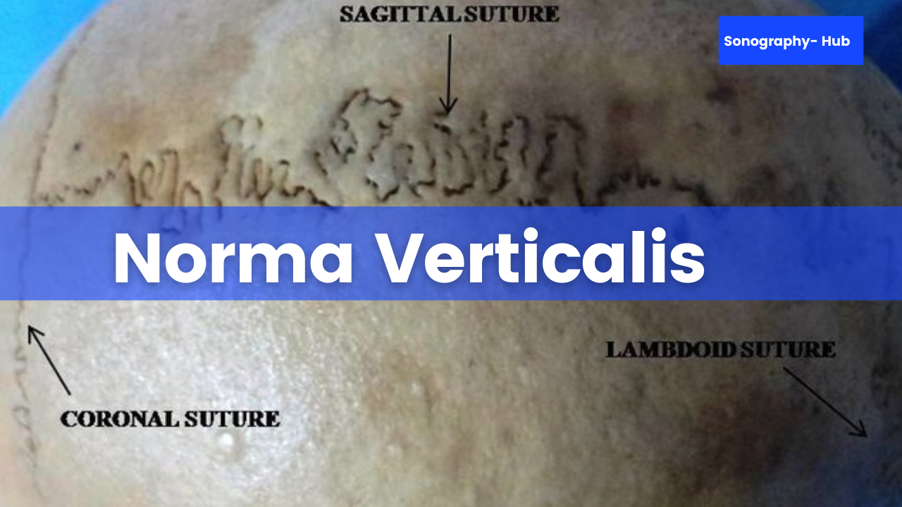
When viewed from above the skull is usually oval in shape. It is wider posteriorly than anteriorly. The shape may be more nearly circular.
Bones saw in Norma Verticalis
Bones that are seen in norma verticalis are:
- The upper part of the frontal bone anteriorly.
- Uppermost part of occipital bone posteriorly.
- A parietal bone on each side.
Sutures
In Norma Verticalis sutures that are seen as following
- Coronal suture
- Sagittal suture
- Lambdoid suture
- Metopic suture (Latin forehead)
1. Coronal Suture
This is placed between the frontal bone and the two parietal bones. The sutures cross the cranial vault from side to side and run downwards and forwards.
2. Sagittal Suture
It is placed in the median plane between the two parietal bones.
3. Lambdoid Suture
It lies posteriorly between the occipital and the two parietal bones, and it runs downwards and forwards across the cranial vault.
4. Metopic Suture
This is occasionally present in about 3 to 8 % of individuals. It lies in the median plane and separated the two halves of the frontal bone, normally, it fuses at 6 years of age.
Clinical Anatomy
Clinical anatomy of Norma Verticalis
- Fontanelles are sites of growth of the skull, permitting the growth of the brain and helped to determine the age.
- In fontanelles fuse early, brain growth is stunted; such children are less intelligent.
- In anterior fontanelle is bulging, there is raised intracranial pressure. If anterior fontanelle is depressed, it shows decreased intracranial pressure, mostly due to dehydration.
- Bones override at the fontanelle helping to decrease the size of the head during vaginal delivery.
- Caput succedaneum is soft tissue swelling on any part of the skull due to rupture of capillaries during delivery. The skull becomes normal within a few days in postnatal life.


Post a Comment