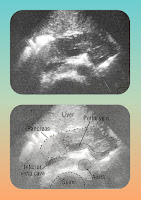
Ultrasound can not distinguish focal pancreatitis from a pancreatic tumor.
Common Errors: when scanning the pancreas, an incorrect diagnosis may be made because:
- The gallbladder is in the midline.
- There are enlarged lymph nodes.
- There is a retroperitoneal mass.
- There is located ascites or an intra-abdominal abscess (including splenic abscess).
- There are hepatic cysts or tumors.
- There are mesenteric cysts.
- There is a hematoma around the duodenum.
- The stomach is partially filled. If the stomach contains fluid, it may resemble a pancreatic cyst; if it contains food, it may mimic a tumor. the adjacent bowel can cause similar errors.
- There are renal cysts, a renal tumor, or a large renal pelvis.
- There is an aortic aneurysm.
- There is an adrenal tumor.
Normal Pancreas & scanning landmarks
The pancreas has about the same echogenicity as the adjacent liver and should appear homogeneous. However, pancreatic echogenicity increases with age. The outline of the normal pancreas is smooth.
When scanning the pancreas, certain anatomical landmarks should be identified, in the following order:
- Aorta
- Inferior vena cava (IVC)
- Superior mesenteric artery
- Splenic vein
- Superior mesenteric vein
- Wall of the stomach
- Common bile duct
The essential landmarks are the superior mesenteric artery and the splenic vein.
What is the normal pancreatic size?
There is great variability in the size and shape of the pancreas. The following guidelines may be helpful.
- The average diameter of the head of the pancreas (A): 2.8 cm.
- The average diameter of the medial part of the body of the pancreas (B): less than 2 cm.
- The average diameter of the tail of the pancreas (C): 2.5 cm.
- The diameter of the pancreatic duct should not exceed 2mm. It is normally smooth, and the wall and the lumen can be identified. The accessory pancreatic duct is seldom visualized.
Small pancreas findings on scanning?
The pancreas is usually smaller in elderly people, but this is not of clinical significance. When there is overall atrophy of the pancreas, the decreases in size is usually uniform throughout the pancreases If there appears to be atrophy of the tail of the pancreas alone (the head appearing normal), Then a tumor in the head of the pancreas must be suspected (fig-1a) The head must be scanned carefully because chronic pancreatitis in the body and tail may be associated with a slow growing-tumour in the head of the pancreas.
If the pancreas is small and irregularly hyperechogenic and non-homogeneous compared with the liver, the cause is usually chronic pancreatitis.
Some basic specifications for small pancreas
Acute pancreatitis may be diffusely enlarged and either normal and hypoechogenic compared with the adjacent liver. The serum amylase is usually elevated, and there is maybe local ileum due to bowel irritation.
When the pancreas is irregular hyperechogenic and diffusely enlarged, there is usually acute pancreatitis superimposed on chronic pancreatitis (fig. 2a).
Diffuse enlargement of the pancreas:
In acute pancreatitis, the pancreas may be diffusely and either normal or hypoechogenic compared with the adjacent liver. The serum amylase is usually elevated. And there may be local ileus due to bowel irritation.
When the pancreas is irregularly hyperechogenic and diffusely enlarged, there's his usual acute pancreatitis superimposed on chronic pancreatitis (Fig. 3a)
What is noncystic?
Focal enlargement is known as noncystic:
Almost all pancreatic tumors are hypoechogenic compared with the normal pancreas.it is not possible to distinguish between focal pancreatitis and pancreatic tumor by ultrasound alone. Even if the serum amylase is elevated. Tumor and pancreatitis can co-exist.


Post a Comment